Cholestatic Vs Hepatocellular Pattern
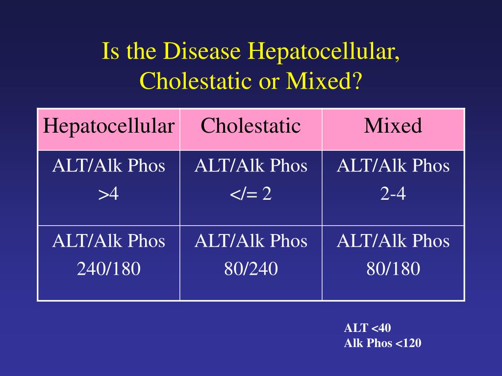
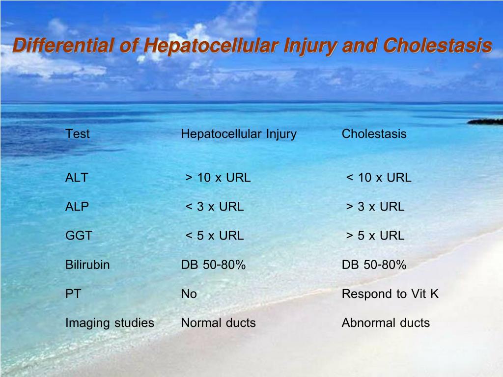
Cholestatic Vs Hepatocellular Pattern - Web there are four major types of liver injury: Web the pattern of alt to alp rise can indicate whether the pathology is primarily cholestatic or hepatocellular: The pattern occurs when there is a disproportionate elevation in alkaline phosphatase (alp) compared to alanine aminotransferase (alt) and aspartate aminotransferase (ast). Web using a schematic approach that classifies enzyme alterations as predominantly hepatocellular or predominantly cholestatic, we review abnormal enzymatic activity within the 2 subgroups, the most common causes of enzyme alteration and suggested initial investigations. Ratio of ast and alt can be useful in differential. Dili is characterized as mixed if the r ratio is between 2 and 5. Alt is more specific for liver damage than ast. Web differentiates cholestatic from hepatocellular liver injury, recommended by acg guidelines. Hepatocellular, autoimmune, cholestatic, and infiltrative (table 1). Web the cholestatic pattern of liver function test abnormalities indicates biliary obstruction. Web an r ratio of greater than 5 defines hepatocellular dili, whereas cholestatic dili is characterized by an r ratio of less than 2. Dili is characterized as mixed if the r ratio is between 2 and 5. Alt is more specific for liver damage than ast. Ratio of ast and alt can be useful in differential. Aminotransferases (ast, alt) generally associated with hepatocellular damage. The pattern occurs when there is a disproportionate elevation in alkaline phosphatase (alp) compared to alanine aminotransferase (alt) and aspartate aminotransferase (ast). Web the three abnormal patterns that can be detected in liver function tests include the hepatocellular pattern, cholestatic pattern, and isolated hyperbilirubinemia pattern, each of which can be acute, subacute, or chronic in presentation. Web there are four major types of liver injury: Generally not associated with cholestasis. Web when both sets of enzymes are elevated, distinguishing between the two patterns of liver disease can be difficult. Web the pattern of alt to alp rise can indicate whether the pathology is primarily cholestatic or hepatocellular: Web when both sets of enzymes are elevated, distinguishing between the two patterns of liver disease can be difficult. Aminotransferases (ast, alt) generally associated with hepatocellular damage. Alt is more specific for liver damage than ast. Web using a schematic approach that. Web the cholestatic pattern of liver function test abnormalities indicates biliary obstruction. Generally not associated with cholestasis. Web overall analysis of liver function tests (lft) transaminitis: The aim of this study was to document the predicted ranges of serum alp values in patients with hepatocellular liver injury and alt or ast values in patients with cholestasis. A hepatocellular pattern is. Web using a schematic approach that classifies enzyme alterations as predominantly hepatocellular or predominantly cholestatic, we review abnormal enzymatic activity within the 2 subgroups, the most common causes of enzyme alteration and suggested initial investigations. The pattern occurs when there is a disproportionate elevation in alkaline phosphatase (alp) compared to alanine aminotransferase (alt) and aspartate aminotransferase (ast). The aim of. Web an r ratio of greater than 5 defines hepatocellular dili, whereas cholestatic dili is characterized by an r ratio of less than 2. Web the pattern of alt to alp rise can indicate whether the pathology is primarily cholestatic or hepatocellular: Ratio of ast and alt can be useful in differential. Web there are four major types of liver. Web the pattern of alt to alp rise can indicate whether the pathology is primarily cholestatic or hepatocellular: Web the cholestatic pattern of liver function test abnormalities indicates biliary obstruction. Aminotransferases (ast, alt) generally associated with hepatocellular damage. Web the three abnormal patterns that can be detected in liver function tests include the hepatocellular pattern, cholestatic pattern, and isolated hyperbilirubinemia. Web the pattern of alt to alp rise can indicate whether the pathology is primarily cholestatic or hepatocellular: Web using a schematic approach that classifies enzyme alterations as predominantly hepatocellular or predominantly cholestatic, we review abnormal enzymatic activity within the 2 subgroups, the most common causes of enzyme alteration and suggested initial investigations. Web the cholestatic pattern of liver function. Ratio of ast and alt can be useful in differential. Web overall analysis of liver function tests (lft) transaminitis: Alt is more specific for liver damage than ast. Web when both sets of enzymes are elevated, distinguishing between the two patterns of liver disease can be difficult. A hepatocellular pattern is marked by isolated or predominant elevations. Web the three abnormal patterns that can be detected in liver function tests include the hepatocellular pattern, cholestatic pattern, and isolated hyperbilirubinemia pattern, each of which can be acute, subacute, or chronic in presentation. The predominant laboratory abnormality defines the pattern of injury. Web using a schematic approach that classifies enzyme alterations as predominantly hepatocellular or predominantly cholestatic, we review. Generally not associated with cholestasis. Web an r ratio of greater than 5 defines hepatocellular dili, whereas cholestatic dili is characterized by an r ratio of less than 2. Aminotransferases (ast, alt) generally associated with hepatocellular damage. Web differentiates cholestatic from hepatocellular liver injury, recommended by acg guidelines. Web using a schematic approach that classifies enzyme alterations as predominantly hepatocellular. The predominant laboratory abnormality defines the pattern of injury. Web the cholestatic pattern of liver function test abnormalities indicates biliary obstruction. Web when both sets of enzymes are elevated, distinguishing between the two patterns of liver disease can be difficult. Web differentiates cholestatic from hepatocellular liver injury, recommended by acg guidelines. Web the three abnormal patterns that can be detected. Web when both sets of enzymes are elevated, distinguishing between the two patterns of liver disease can be difficult. Generally not associated with cholestasis. Web the cholestatic pattern of liver function test abnormalities indicates biliary obstruction. The predominant laboratory abnormality defines the pattern of injury. The pattern occurs when there is a disproportionate elevation in alkaline phosphatase (alp) compared to alanine aminotransferase (alt) and aspartate aminotransferase (ast). Alt is more specific for liver damage than ast. Web an r ratio of greater than 5 defines hepatocellular dili, whereas cholestatic dili is characterized by an r ratio of less than 2. Hepatocellular, autoimmune, cholestatic, and infiltrative (table 1). Ratio of ast and alt can be useful in differential. Dili is characterized as mixed if the r ratio is between 2 and 5. Web overall analysis of liver function tests (lft) transaminitis: Web the pattern of alt to alp rise can indicate whether the pathology is primarily cholestatic or hepatocellular: Web the three abnormal patterns that can be detected in liver function tests include the hepatocellular pattern, cholestatic pattern, and isolated hyperbilirubinemia pattern, each of which can be acute, subacute, or chronic in presentation. Aminotransferases (ast, alt) generally associated with hepatocellular damage. Web using a schematic approach that classifies enzyme alterations as predominantly hepatocellular or predominantly cholestatic, we review abnormal enzymatic activity within the 2 subgroups, the most common causes of enzyme alteration and suggested initial investigations.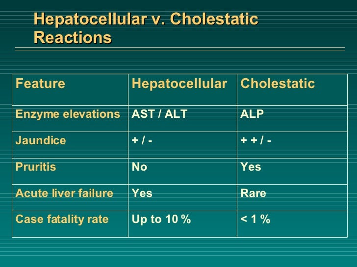
Liver Failure Case

Liver Histology Clinics in Liver Disease

Laboratory Associations with Hepatocellular and Cholestatic Patterns of

PPT Abnormal LFTs PowerPoint Presentation, free download ID139175
Liver function tests in primary care bpacnz
PPT Work up of the Asymptomatic Patient with Liver Enzyme
PPT Liver Function Test s PowerPoint Presentation, free download ID

Pin on Infographics
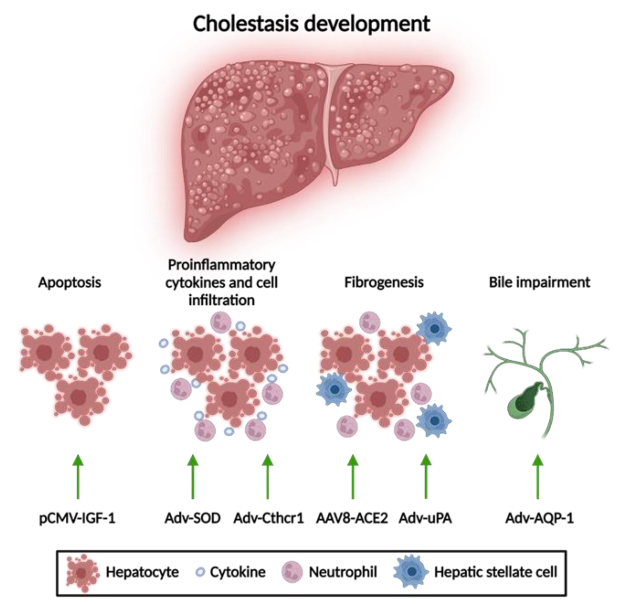
Gene Therapy for Cholestasis Encyclopedia MDPI

Liver Enzymes (hepatic vs cholestatic patterns) Sketchy Medicine
Web Differentiates Cholestatic From Hepatocellular Liver Injury, Recommended By Acg Guidelines.
The Aim Of This Study Was To Document The Predicted Ranges Of Serum Alp Values In Patients With Hepatocellular Liver Injury And Alt Or Ast Values In Patients With Cholestasis.
A Hepatocellular Pattern Is Marked By Isolated Or Predominant Elevations.
Web There Are Four Major Types Of Liver Injury:
Related Post: